


What is cataract? – A cataract is a clouding of the lens of the eye or in its envelope (lens capsule), varying in degree from slight to complete opacity, which results in a decreased vision or complete blindness.
What are the causes of cataracts? – Cataracts in dogs most frequently develop as a result of diabetes (mellitus). Almost 90% of diabetic dogs develop cataracts within the first 12-18 months after being diagnosed with a disease. In many canine breeds, cataracts are hereditary (genetic). Some dogs are born with hereditary cataracts (congenital cataracts), while some dogs develop cataracts as they are aging. Cataracts can also be caused by ocular trauma and inflammation.
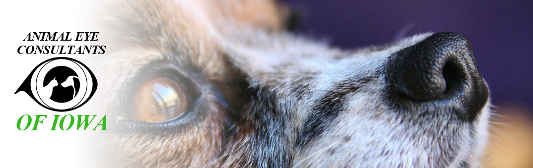
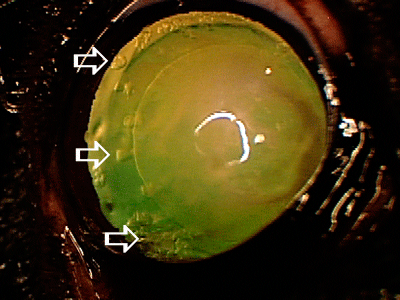
 Mature diabetic cataract in a Bichon. Cataract is abnormal change of the lens material resulting in the white milky appearance (arrow). Sometimes diabetic cataracts have rapid progression over the course of several weeks or even days resulting in sudden onset of vision loss. Rapidly progressive cataracts can be very dangerous due to inflammation and irritation they can cause, and in some cases may result in the acute glaucoma development. If you detect sudden onset of vision loss or white eye milky appearance, contact your veterinarian immediately so eye can be evaluated.
Should I be concerned if my pet is diagnosed with cataracts? Regardless of the cause of cataracts, it is very important to identify and monitor them from the very early stage of development. Lenses affected by cataracts are continuously leaking lens material, which can be extremely irritating and dangerous to the eye. Leaking lens material is recognized by the immune system as a foreign material, and aggressive auto-immune reactions can develop, resulting in the pain, ocular discomfort, blindness, and ultimately loss of the eye as a result of the untreated cataracts.
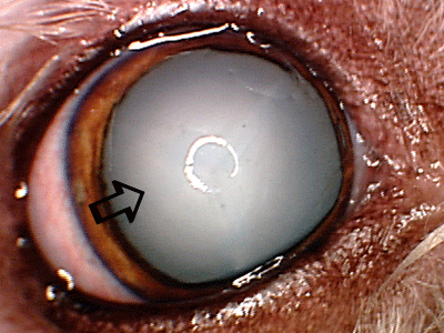
 Cataract complications – chronic presence of cataracts may result in many complications resulting in the pain, discomfort and eventual need for the surgical eye removal. This patient had long standing cataract (white arrow), which resulted in glaucoma (elevation of intraocular pressure) resulting in ocular pain, redness (black arrow) and eventual need for the surgical eye removal.
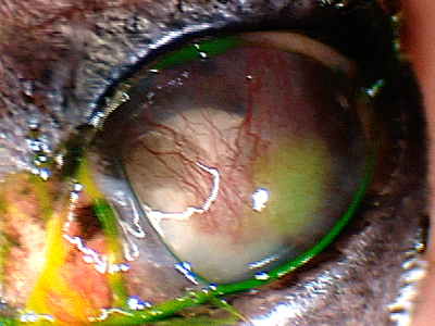 Cataract complications – Chronic presence of cataract in this patient resulted in chronic inflammation, glaucoma, development of corneal ulcer and ultimate need for the surgical eye removal.
 Small hereditary cataract in a mixed breed dog. These cataracts usually do not progress, and they rarely cause any vision problems or ocular irritation. Routine annual evaluation of the cataract progression should be pursued with your veterinarian.
Small cataracts, which are not resulting in the significant ocular irritation or vision disturbances, are usually just monitored. Cataracts which cause significant vision problems or ocular irritation must be surgically removed to restore the vision and reduce the risk of the possible loss of the eye associated with uncontrolled inflammation in the eye. Because of numerous complications associated with cataracts, it is important to medically treat and monitor cataracts in all cases where surgery is not performed. Medical treatments usually include topical anti-inflammatory eye drops applied once or twice daily for life. In some cases systemic anti-inflammatory medications may be needed.
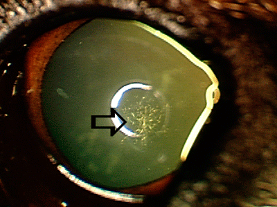
 Early cataracts (white arrows). These early cataracts do not interfere with vision, however rapid progression of cataracts is possible, which may result in vision loss and different ocular complications. Regular monitoring of cataract progression should be pursued with your local veterinarian.
Is there a treatment for cataracts? Cataract surgery is a routine procedure performed by veterinary ophthalmologists, which has 95% success rate. While the initial financial investment for this type of surgery is high ($2800-$3500), almost identical amount of money is usually spent (over the period of 3-5 years) for medical and surgical treatment of eyes with cataracts where cataract surgery was not pursued, and complications eventually developed. Pursuing cataract surgery reverses blindness and ultimately dramatically reduces chances of long-term complications, pain and loss of eye usually seen in eyes with untreated cataracts.
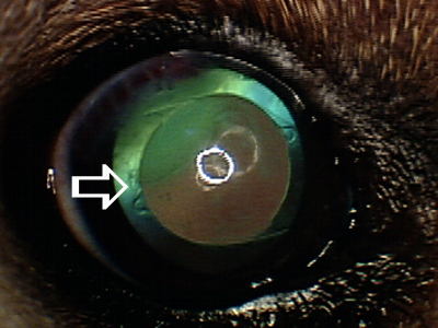 Cataract surgery is effective method for permanently removing cataracts and restoring normal vision. Artificial lens (white arrow) is usually placed in the eye with a goal of correcting vision to the normal levels. Removal of cataracts eliminates risks of chronic cataract-induced inflammation, and associated complications, which may result in blindness and loss of eye (retinal detachment, glaucoma, intraocular hemorrhage).
What are the possible complications of cataract surgery?The most frequent complications of cataract surgery are glaucoma (development of the high intraocular pressure in the eye), retinal detachment, and severe intraocular inflammation or infection. These complications can resolve with aggressive medical treatment, however in some cases additional surgical procedure may be needed to provide good visual outcome. In very rare cases, eye cannot be saved and surgical removal of the needs to be pursued. Fortunately, these complications are relatively rare.
What can be done to prevent surgical complications associated with cataract surgery? Your ophthalmologist will perform detailed evaluation of the eye prior to cataract surgery to identify any possible problems (intraocular inflammation, glaucoma predisposition, possible abnormality in retinal function and structure). It is very important that dental cleaning is done so possible intraocular infection due to dental bacteria can be prevented after cataract surgery. Many diabetic dogs have urinary tract infections due to the presence of sugar in the urine, so starting systemic antibiotics before cataract surgery in dogs with urinary tract infection is also an excellent strategy to prevent complications after cataract surgery.
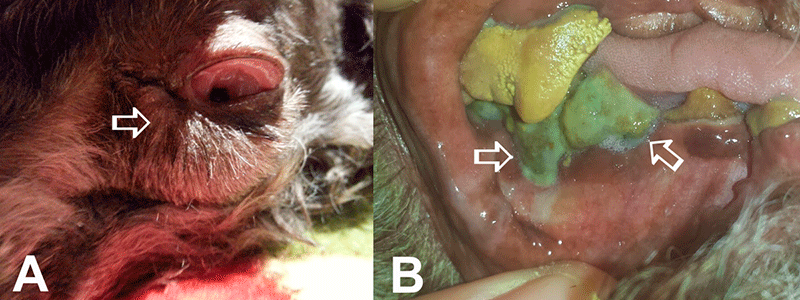
Postoperative complications after cataract surgery. A) Severe postoperative swelling of ocular and periocular structures (arrow) due to bad teeth was present in this patient, which developed one day after emergency lens removal surgery. B) Dental images from the same patient showed extensive dental tartar presence with inflammation and infection of gingiva (arrows). VISIT YOUR LOCAL VETERINARIAN AND DISCUSS DENTAL CLEANING PRIOR TO PURSUING CATARACT SURGERY!
Does cataract surgery have to be repeated? Cataract surgery is done just once and at that time lens material affected by cataract is completely removed and replaced with an artificial plastic lens, which stays in the eye permanently, so there is no need to repeat cataract surgery again.
Is there a problem if artificial lens is not placed in the eye during cataract surgery? In some cases an artificial plastic lens can’t be placed in the eye, which may result in decreased performance in some dogs, while majority of dogs do have almost normal functional vision even without placement of artificial lenses. Dogs without artificial lenses usually have problems seeing small objects in a close proximity to the head.
I can’t afford cataract surgery – what are my options? Cataract surgery requires significant financial commitment, and if resources are not available to pursue cataract surgery, topical (and in some cases systemic) anti-inflammatory therapy needs to be pursued. Unfortunately, cataract progression always results in numerous complications (chronic inflammation, lens luxation, retinal detachment, intraocular bleeding, secondary glaucoma), which ultimately dictate a need for a surgical eye removal. Cataract is a surgical disease, and there has to be a clear understanding that surgical eye removal will likely be needed in the future, if cataract surgery repair is not an option.
»back to top
ACVO Cataract Video - Click Here
Corneal Ulcers – Corneal ulcer (ulcerative keratitis) is a condition characterized by a damage to one or multiple corneal layers. The cornea is the transparent structure at the front of the eye, which allows for images to be projected to the neurosensory part of the eye (retina). Cornea does not have blood vessels and strictly depends upon the oxygen supply from tears and intraocular fluid (aqueous humor). Due to its delicate structure (thickness is 0.5 mm), cornea is very sensitive to the trauma and inadequate oxygenation. Since numerous bacteria normally live on the corneal surface, any damage to the corneal structure may result in the rapid bacterial infection, ultimately leading to the corneal destruction. Corneal ulcers may be shallow, involving the most superficial layers of the cornea or may be very deep, damaging nearly all of the corneal layers. Shallow ulcers are usually treated medically, while deep corneal ulcers may require surgery to rebuild the corneal integrity.
Shallow ulcers normally heal within 5-7 days. If complete ulcer healing is not present within 7-10 days, these ulcers become non-healing or indolent ulcers. Indolent ulcers can't heal with the use of medical therapy only, and high speed corneal burring or needle scrapping of the ulcer surface is usually performed to stimulate the ulcer healing. This procedure is usually done with the use of topical anesthetic drops or general sedative agents.
The most frequent cause for corneal ulcers is the poor quality of quantity of tears, which is frequently present in dogs with seasonal or food allergies affecting the eyes, eyelids and skin in general. Abnormal shape of eyelids and foreign bodies are also among causes of corneal ulcers in dogs. Symptoms of corneal ulcers usually include squinting, excessive eye redness and discharge, sensitivity to light and corneal cloudiness (hazy or blue color).
If recognized early, most corneal ulcers will heal quickly with the aggressive medical therapy. Deep ulcers are almost always infected and may require hospitalization for hourly medical treatment for several days. Corneal ulcers which destroy more than 60-70% of corneal structure, usually require surgical treatment with a goal or restoring corneal integrity and preventing the loss of the eye. It is very important that rubbing of injured eyes is prevented with the use of an Elizabethan plastic protective collar. Excessive rubbing on injured eye may make more damage to the cornea and bring more of bacteria from paws to the eye.
»back to top
Dry Eye Disease (KCS - Keratoconjunctivitis Sicca) – KCS is a frequently encountered condition in dogs, which develops as a result of the decreased quality or quantity of the tear film (abnormally low tear production).
The tear film covers the surface of the eye (cornea), and is comprised of 3 layers (water, oil, and mucous). These layers of tear film are produced by lacrimal glands, eyelid skin and conjunctival cells (conjunctiva is the white tissue surrounding the eye). Any condition which affects these structures may result in changes of the tear film quality. Tear film abnormalities are very frequently encountered in dogs with seasonal and food allergies, so pets with these conditions and "red eye" appearance should have a thorough ophthalmology evaluation.
Since tear film provides nutrients, anti-microbial protection and oxygen to the surface of the eye, lack of tears may result in the excessive irritation, discomfort, development of corneal ulcers, or chronic scarring of the cornea resulting in impaired vision or even blindness. Modern medical treatments are very effective in terms of controlling dry eye disease clinical symptoms. Some patients do not respond to medications, and a specific surgical procedure (parotid duct transposition) can be performed. Parotid duct transposition results in the transposition of the salivary duct to the lower eyelid of affected eyes, so saliva can moisturize the eye surface.
»back to top
Glaucoma – Glaucoma is disease characterized by the high elevation of pressure inside the eye (intraocular pressure), which results in a damage to the eye and eventually blindness. Glaucoma is a frequent cause of blindness in our pets, and most frequently is a hereditary disease affecting middle-aged dogs.
The eye shape is maintained by the intraocular fluid (aqueous humor), which is continuously produced and drained from the eye. Similar to the open faucet and drain, the system is well maintained as long as the drain system in functional. If drain systems of the eye get plugged, the intraocular fluid will start to accumulate in the eye resulting in the rise of pressure. This build-up of the high pressure in the eye can damage many eye structures in the very short period of time.
Medical and surgical therapies for glaucoma can reduce production of the intraocular fluid ("closing the faucet") and increase the flow of the intraocular fluid from the eye ("unplugging the drain") with a goal to maintain the intraocular pressure of 15-20 mmHg.
Detailed glaucoma screening is recommended for all breeding dogs, and also for pets with a history of disease in their family. Early detection and treatment of glaucoma may dramatically delay the blindness associated with this disease.
»back to top
Eyelid abnormalities – Eyelids are the primary protective structure to the ocular surface. Eyelid abnormalities resulting in the abnormal eyelid function or ocular surface irritation may result in development of corneal ulcers or scar/pigmented tissue, ultimately resulting in decreased vision or even blindness. Eyelid abnormalities can be effectively corrected with different surgical techniques.
• Entropion is an inward rolling of the eyelids that may involve the upper lids, lower lids, or both. This condition creates ocular pain and discomfort, eye redness, excessive tearing and if not repaired corneal ulcers and scarring. Surgery is usually corrective. However, in very young dogs temporary "tacking" (placement of eyelid sutures to roll out the eyelid margin away from the cornea) can be done. In some breeds (Shar Pei, Chow-Chow) more extensive surgery needs to be done to correct the entropion.
• Ectropion is an outward rolling of the eyelids. It can lead to chronic irritation, excessive tear evaporation and ocular discharge. Frequently only eye lubrication is sufficient to control the ocular discomfort. However, surgical correction may be necessary in some cases.
• Eyelid Masses can cause ocular irritation, discomfort, and corneal scarring. Most eyelid masses in dogs are benign and cured by surgical removal, but some eyelid masses are malignant in nature and may require aggressive medical and surgical therapy. Fortunately, eyelid tumors in dogs rarely spread to the rest of the body.
• Prolapsed third eyelid gland ("cherry eye") is a condition where the lacrimal gland of the third eyelid prolapses from its normal position, resulting in the swelling and decreased function. This condition can cause ocular irritation, discomfort, decreased tear production and corneal scarring. Prolapsed third eyelid glands must be preserved and repositioned, so normal tear production can be secured.
»back to top
Sudden onset of blindness – Numerous ocular and systemic diseases can cause the sudden onset of blindness in dogs. Fortunately, many of these conditions are reversible and easily treated if diagnosed in the early stage of disease. Any sudden decline in vision should be immediately evaluated by your veterinarian or ophthalmologist.
Sudden onset of blindness and "red eye" appearance - most frequent conditions causing excessive eye redness and blindness are glaucoma, lens luxation, severe intraocular inflammation (uveitis, chorioretinitis), acute trauma, bleeding disorders resulting in the blood accumulation inside the eye, and rapid cataract progression (most frequently seen in diabetic dogs, as a result of the trauma or in some forms of hereditary cataracts - Bichon Frise breed).
Sudden onset of blindness and "normal" eye appearance - most frequent conditions causing the sudden onset of blindness and normal eye appearance are sudden acquired retinal degeneration syndrome (SARDs), immune-mediated retinitis (IMR), retinal detachments, optic neuritis/meningitis, toxin exposure (particularly ivermectin exposure), optic nerve tumors, orbital tumors, brain tumors, ischemic events (stroke) affecting brain regions responsible for vision processing.
Many conditions resulting in sudden onset of blindness are result of the systemic diseases, which can be life threatening. Careful systemic examination of patients with a sudden onset of blindness is a must, in order to prevent more serious consequences for the systemic health.
»back to top

