Basic physiological principles of the chromatic pupil light reflex testing.
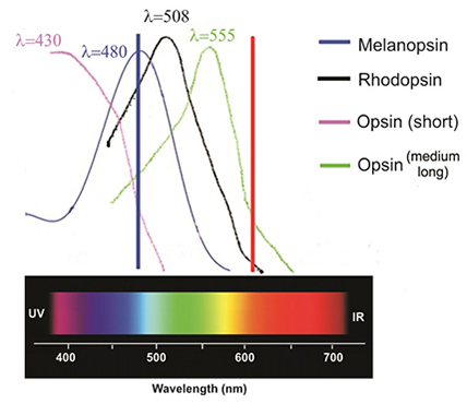
Physiological principle of cPLR testing in canine eyes (image is provided by Dr Sinisa Grozdanic, DVM, PhD, Dipl ACVO). Blue light of 480 nm wavelength activates short and medium-long wave length cone opsins, melanopsin and rhodopsin. The red light of 630 nm wavelength can’t activate short wave length cone opsin and melanopsin. Since red light of 630 nm wavelength is activating the far edge of the rhodopsin and medium-long wave cone opsin sensitivity spectrum, pathological change in these specific photoreceptors (rods and medium-long wavelength cones which are dominant photoreceptors in the canine retina) may result in abnormal electrical response, which can be evident as an incomplete PLR or PLR with a presence of pupil escape, particularly after the red light illumination.
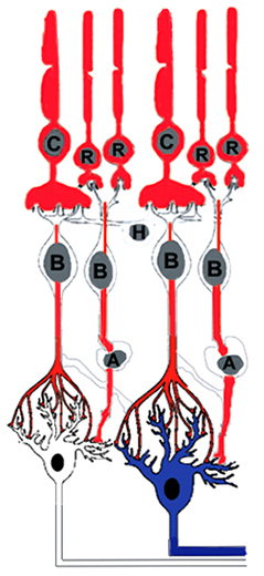
Principle of melanopsin and non-melanopsin mediated PLR activation. Red light stimulus activates the rod-cone pathway. Any pathology in the rods, cones and/or inner retinal neurons connecting to ipRGCs (blue neurons) results in decreased PLR response to red light. Optic nerve lesions effectively impair signal transmission (melanopsin and non-melanopsin mediated) regardless of the type of light stimulus used for testing. In patients who recover from the optic neuritis, damage to ipRGCs may prevent recovery of PLR and dazzle response. However, if the sufficient number of RGCs (white cells) remains, vision can still be processed. C-cone; R-rod; H-horizontal cell, B-bipolar cell, A-amacrine cell.
Scientific Partners
Vision Biomedical SolutionsNikola Tesla 115, Apatin 25260, Serbia
website: www.cpupil.com
email: cpupilreflex@gmail.com
phone: 21105503